Cells 2012
Cells 2012 kdorfman Tue, 07/03/2012 - 17:26Lab 1: FL microscope
Lab 1: FL microscope kdorfman Mon, 07/30/2012 - 18:0310 slides w/ DAPI
(1 per microscope + 2 extra)
Micrometers
Lab 2: Organelles
Lab 2: Organelles kdorfman Mon, 07/30/2012 - 18:265 or 6 slides each:
- Rhodamine Phalloidin "1G"
- Stained for rat anti-tubulin + goat anti-rat FITC (or GFP-tubulin cells) "2B"
- Acridine orange stained for nucleoli "3G"
- Stained with with mouse anti-Golgi + goat anti-mouse 568 "4G" (formaldehyde fixed)
- Stained with rabbit anti-ZO1 + goat anti-rabbit Cy (cell-junction) "5G"
Lab 3: live cells; staining
Lab 3: live cells; staining kdorfman Mon, 07/30/2012 - 18:3120 fixed coverslips for phalloidin staining
3 MatTeks per group for mitotracker and ?
aliquots of
- phalloidin
- mitotracker
- non-CO2 medium
Lab 4 - discussion
Lab 4 - discussion kdorfman Mon, 07/30/2012 - 20:15Finish looking at slides, discuss cell division
Lab 5: mitosis
Lab 5: mitosis kdorfman Mon, 07/30/2012 - 20:1810 slides DAPI + tubulin
2 MatTeks per group each:
- LLCPK with GFP tubulin
- LLCPK parentals
NUC-Blue for staining chromosomes in live cells (Invitrogen R37605 or Fisher NC0291762), 1 bottle per station
- Remove from culture dish after ~10 minutes. Replace stain medium with fresh medium. Otherwise it gets brighter and brighter as movie goes on
Non-CO2 medium
Lab 6: Mitosis
Lab 6: Mitosis kdorfman Mon, 07/30/2012 - 20:454 MatTeks per group of LL-GFP-tubulin
Double-strength inhibitors, made up in non-CO2 medium:
STLC Sigma 164739 (Fisher 50-703-1833), MW = 363.47
need 1 mL aliquots 10 µM in non-CO2 medium for students
Make 100 mM stock:
- 1 g in bottle.
- Mix with 27.5 mL DMSO
- heat to 65C (~1 hr), vortex
- if necessary, filter sterilize to remove insoluble particles.
- save as 1 mL aliquots. (dilute 1:10,000 to use) (dilute 1:100 to make 1 mM)
Make 1 mM stock from the 100 mM stock
- 10 µL 1mM stock
- 990 µL DMSO
- vortex
- it will crystallize in the refrigerator. Warm and vortex to redissolve it.
Make 17 mL 10 µM in medium for students
- 170 µL 1 mM stock
- medium to 17 mL
- aliquot
Make 10 µL aliquots of 1 mM stock
- label says to add 990 µL medium to make 10 µM working solution
from Alyssa:madke STLC in DMSO and that she heated to 42C, and vortexed. She says it went from clear to yellow/brown. we use the reagent at micromolar - 1-10 µM. Her stock solution was 1mM.
students will be adding 1 mL of inhibitor to 1 mL medium in dish; the ready-to-use 1 mL (in medium) should be at least 2µM
- Taxol (20 µM final)
- soluble in DMSO, not water.
- students pick either Taxol or nocodazole, so make only 10 each
- each tube has 10 µL 10 mM (if it precipitates, add 10 uL DMSO and vortex, then add 480 uL medium or buffer)
- add 490 µL to make 200µM in two tubes!
- mix with medium in 15 mL conical to make 10 mL at 20 µM
Nocodazole (3.3 µM final)
- each tube has 3 µL 33mM
- add 97 µL to tube to make 100 µL of 1 mM
- 33 µL 1mM + 10 mL medium = 3.3 µM
Nuc Blue from Invitrogen (or Fisher NC0291762), 1 bottle per station
- Remove from culture dish after ~10 minutes. Replace stain medium with fresh medium. Otherwise it gets brighter and brighter as movie goes on
Lab 7: Cytokinesis
Lab 7: Cytokinesis kdorfman Mon, 07/30/2012 - 20:58Myosin & tubulin expressing cells
2 MatTeks each per group
For ML-7, the final conc should be 75uM, so the 2X needs to be 150uM. For the Jasplak: the final conc should be 7uM so the 2X needs to be 14uM (but if 15 is easier, go with it). we use latrunculin at 5uM, and I think cytoD should be similar (I think I previously gave a range for cytoD of 1-10uM). Let's go with 5, so the 2X would be 10uM.
Inhibitors in non-CO2 medium (1 mL aliquots) each group does one (so make 5 of each):
cytochalasin D Sigma C2618 or C8273
- tubes have 40 µL 2.5 mM
- label says add 460 µL to make 500 µL of 200 µM
- need 10 µM (2x final working concentration)
- 20 µL 2.5 mM
- 5 mL non0CO2 medium
- gives 5 1 mL aliquots at 10 µM
Jasplakinolide – Santa Cruz biochemical, product sc202191 F
- inducer of actin polymerization & stabilization
- F-actin probe
- MW = ~710
- powder is stable frozen over a year
- stock is stable 3-4 mo at -20C
- Make 1 mM stock with 50 µg in 70 µL DMSO
- Add 5 mL non-CO2 medium to make ~14 µM
- 50 nM to 5 μM working range
- minutes to hours incubation time
ML-7 Sigma, 12764.
- soluble 10mg/mL in 50% EtOH
- MW = 452.74
- 5 mg in bottle
- add 1100 µL to make 10 mM solution
- used at ~50 µM (1 µL/1mL) (Pat says 75 µM)
- 75 µL 10 mM + 5 mL non-CO2 medium makes 150 µM
- make 15 µL aliquots of 10 mM. Add 985 µL medium to make 150 µM
Lab 8: projects
Lab 8: projects kdorfman Mon, 07/30/2012 - 21:04Students limited to 3 MatTeks of one of the following:
- GFP tubulin
- tubulin-myosin cells
Plus their choice of inhibitors:
- latrunculin
- taxol
- nocodazole
- jasplakinolide
- ML-7
- STLC
Lab 9: Reports
Lab 9: Reports kdorfman Mon, 07/30/2012 - 21:08Report on mitosis experiments
RNA
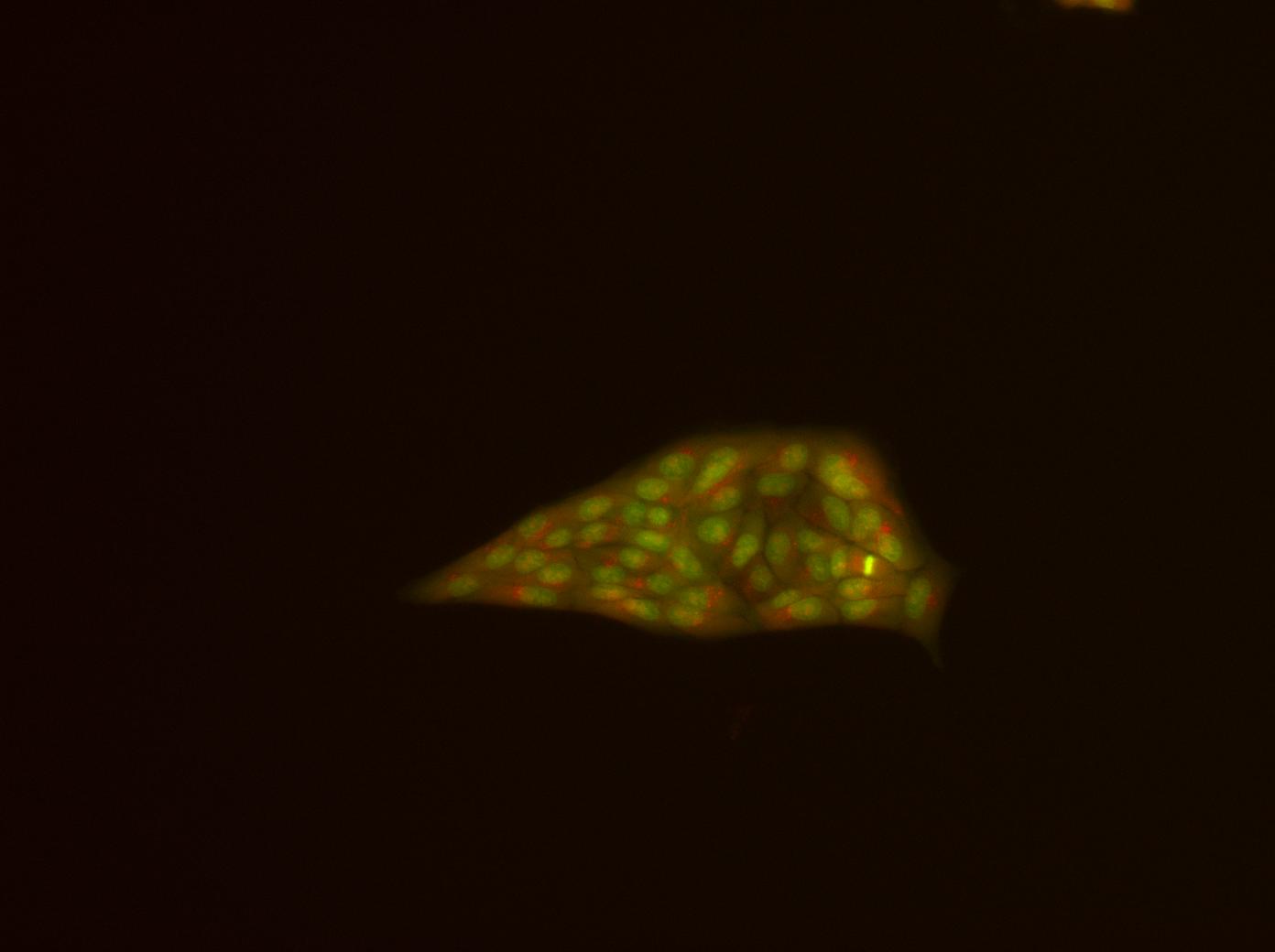
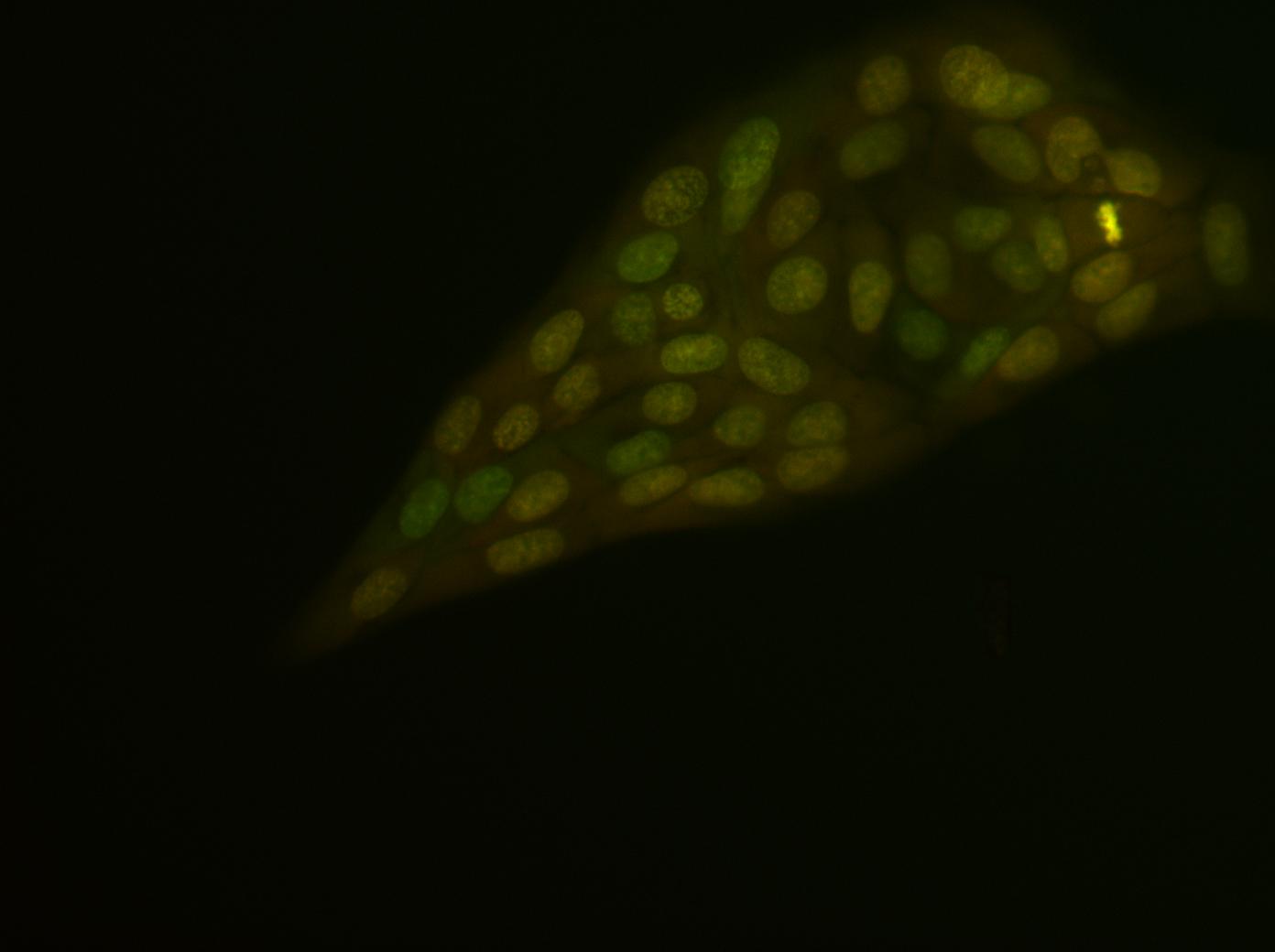
RNA kdorfman Tue, 07/03/2012 - 17:40Acridine orange
Acridine orange kdorfman Fri, 08/03/2012 - 21:01Sigma A8097-10 mL
10 mg/mL = 33mM
MW = 301.81
Use at ~20µM
1.2 µL added to 2 mL in dish
incubate 15 min at 37
change medium
incubate 15 min at 37
Replace medium with PBS
observe with
- B excite - G emission: dsDNA
- G excite - R emission: RNA, ssDNA
Got good results initially, but within minutes, cells started to ball up and pull off the substrate. Will try new PBS first, then a concentration gradient of acridine orange.
Old PBS produced fast shrinking and balling up. Newer PBS was less bad. Now try with non-CO2 medium. Does it interfere with fluorescence?
No cell shrinkage in non-CO2 medium. At short exposure times, there is a background glow, but picking the right exposure takes care of that.
This is time sensitive. The red emission (which should be RNA & ssDNA) gradually fades, or overlies the green emmision (which should be ds DNA)
See attached images, taken in order 1, 2, 3. #3 is at about 15 minutes. 1 & 2 are 100x, 3 is 200x.
SYTO RNASelec
SYTO RNASelec kdorfman Fri, 08/03/2012 - 20:59SYTO RNASelect Green Fluorescent Cell Stain (Invitrogen S32703)
$210 from Invitrogen
Fluoresces green when bound to RNA.
excite: 490 nm, emit: 530 nm
Can be used in live or fixed cells. Fix in methanol, NOT formaldehyde!
Don't use in conjunction with red-orange dyes.
Stock:
100µL 5 mM in DMSO. Store at -20C, dessicated, dark.
To thaw: warm to RT, spin down.
Should be stable for >= 1 year.
Make labeling solution
Make 5µM intermediate stock:
2 µL 5mM stock + 1998 µL medium or PBS
Make 20 100 µL aliquots in 1.5 mL tubes
Label: RNASelect - 100 µL - 5 µM intermediate stock in PBS; Add 900 µL to make labeling solution
Make 500 nM labeling solution in medium or PBS
100 µL 5 µM intermediate + 900 µL medium
Protocol
Live cells
- Cells on coverslip
- Warm 500 nM labeling solution to 37C
- Incubate at 37C 20 min
- Rinse twice in PBS or medium
- Add warm medium, let cells rest 5 min
- Fix in chilled methanol 10 min at -20C
- Several washes in PBS
Fixed cells
- Remove coverslip from medium
- Fix in chilled methanol 10 min at -20C
- Remove methanol, let slip sit in PBS 5 min
- Apply labeling solution 20 min RT
- Wash 5 min in PBS
- Mount coverslip