Counting cells
Counting cells kdorfman Thu, 09/12/2019 - 16:58Just before plating, after cold medium has been added to the trypsinized cells,
mix 0.1 mL cell suspension with 0.1 mL 0.4% trypan blue. Live cells will not take up trypan blue. (Use a different dilution factor if cells are uncountable)
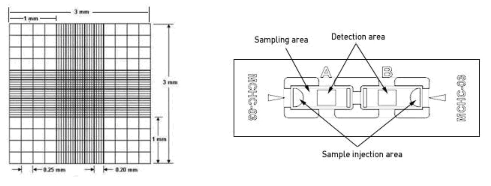
inject 10 uL to one side of the hemocytometer (see image attached below).
Inspect at 10x.
Count the number of live (clear) cells in all 9 squares of the hemocyometer. (Volume = 3mm * 3 mm * 0.1 mm = 0.9 uL) (Count in 4 1mm x 1mm squares if cells are very numerous)
Calculate the live cell concentration (# live cells * 1.11)/1uL for 9 squares,
(# live cells * 2.5/uL for 4 squares)correct for dilution factor
Estimating cell numbers
Estimating cell numbers kdorfman Tue, 08/22/2023 - 17:23From Useful Numbers for Cell culture
(See also counting cells)
| Container | Surface area (cm2) | Seeding density* | Cells at confluency | mL growth medium |
|---|---|---|---|---|
| 35 mm dish | 8.8 | 0.3 x 10⁶ | 1.2 x 10⁶ | 2 |
| 60 mm dish | 21.5 | 0.8 x 10⁶ | 3.2 x 10⁶ | 5 |
| 100 mm dish | 56.7 | 2.2 x 10⁶ | 8.8 x 10⁶ | 12 |
| 150 mm dish | 145 | 5.0 x 10⁶ | 20.0 x 10⁶ | 30 |
| 6-well plate | 9.6 | 0.3 x 10⁶ | 1.2 x 10⁶ | 1 to 3 |
| 12-well plate (~20 mm) | 3.5 | 0.1 x 10⁶ | 0.5 x 10⁶ | 1 to 2 |
| 24-well plate (~15 mm) | 1.9 | 0.05 x 10⁶ | 0.24 x 10⁶ | 0.5 to 1.0 |
| 48-well plate | 1.1 | 0.03 x 10⁶ | 0.12 x 10⁶ | 0.2 to 0.4 |
| 96-well plate | 0.32 | 0.01 106 | 0.04 x 10⁶ | 0.1 to 0.2 |
| T-12.5 flask | 12.5 | 0.35 x 10⁶ | 1.4 x 10⁶ | 1.5-2.5 |
| T-25 flask | 25 | 0.7 x 10⁶ | 2.8 x 10⁶ | 3–5 |
| T-75 flask | 75 | 2.1 x 10⁶ | 8.4 x 10⁶ | 8–15 |
| T-175 flask | 175 | 4.9 x 10⁶ | 23.3 x 10⁶ | 35–53 |
| T-225 flask | 225 | 6.3 x 10⁶ | 30 x 106 | 45–68 |
- Seeding density is given for each culture vessel type as follows:
- Dishes and Flasks: Cells per vessel;
- Culture plates: Cells per well
Hemocytometer Cautions
Hemocytometer Cautions kdorfman Fri, 05/29/2020 - 13:11Use of the Hemacytometer for the Determination of Cell Numbers
Counting cells by the use of a hemacytometer is a convenient and practical method of determining cell numbers in the case that the Coulter counter is out-of-order temporarily. (It is not that bad.) The hemacytometer consists of two chambers, each of which is divided into nine 1.0 mm squares. A cover glass is supported 0.1 mm over these squares so that the total volume over each square is 1.0 mm x 0.1 mm or 0.1 mm3, or 10-4 cm3. Since 1 cm3 is approximately equivalent to 1 ml, the cell concentration per ml will be the average count per square x 104.
Hemacytometer counts are subject to the following sources of error:
- 1. Unequal cell distribution in the sample
- 2. Improper filling of chambers (too much or too little)
- 3. Failure to adopt a convention for counting cells in contact with the boundaries lines or with each other (be consistent)
- 4. Statistical error
With careful attention to detail, the overall error can be reduced to about 15%. It is assumed that the total volume in the chamber represents a random sample. This will not be a valid assumption unless the suspension consists of individual well-separated cells. Cell distribution in the hemacytometer chamber depends on the particle number, not particle mass. Thus, cell clumps will distribute in the same way as single cells and can distort the result. Unless 90% or more of the cells are free from contact with other cells, the count should be repeated with a new sample. A sample will not be representative if the cells are allowed to settle before a sample is taken. Always mix the cell suspension thoroughly before sampling. The cell suspension should be diluted so that each such square has between 20 - 50 cells (2-5 x 10 5 cells/ml). A total of 300 - 400 cells should be counted, since the counting error is approximated by the square root of the total count. A common convention is to count cells that touch the middle lines (of the triple lines) to the left and top of the square, but do not count cells similarly located to the right and bottom. Hemacytometer counts do not distinguish between living and dead cells. A number of stains are useful to make this distinction. Trypan blue among others (Erythrosin B, Nigrosin) can be used: the nuclei of damaged or dead cells take up the stain. If more than 20% of the nuclei are stained, the result is probably significant. Although the trypan stain distinction has been questioned, it is simple and gives a good approximation. See procedure here
References:
From the Laboratory of Dr. Allan Bradley
Baylor College of Medicine, Houston, Texas